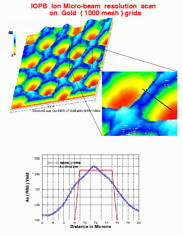
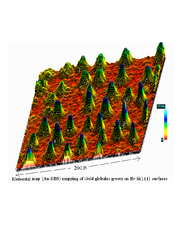
An early resolution scan on 1000 mesh Au grid and an Au elemental map of Au islands sitting on a Si(111) surface are shown as typical examples.
D.P.Mahapatra Last modified: Mon Apr 28 17:26:01 IST 2008
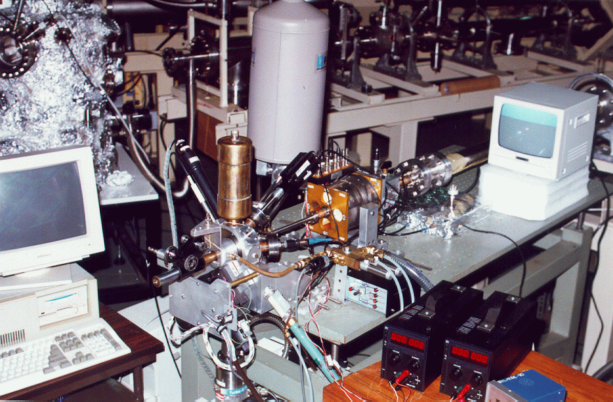
An ion micro-beam facility has been developed at the Institute
in collaboration with
SUNY at Albany (Indo-US
Collaboration). It has been developed along the -30 deg port
of the switching magnet. The system has a micro-slit at the
beam focus following the swiching magnet. This is always fixed
at 100 micron. This is followed by a 5 m section of drift tube
at the end of which lies a micro-quad (Dyer Enegy, USA). The
system has a demagnification ratio of 15:1. The IOP microbeam
system had been designed for a 0.5 micron beam spot. The
detectors include: a) an SEM detector, b) an X-ray detector
(Si(Li)), c) a Si surface barrier detector, (all in front), and
d) an optical microscope at the back for beam focusing and
positioning. The system is capable of generating x-ray as
well as charge particle images of a sample ~ 100 micron square
in area. In some earlier runs we had gone to about 3 micron
in beam size which has been sufficient to get some interesting
pictures of Au and Ag islands on Si (111) surface, x-ray image
of a SQUID structure. Some attempts had also been made to use
a proton beam for writing purposes on plymers etc.
(Collaborators: D. P. Mahapatra, B. N. Dev, B. Rout, H. Bakhru,
A. Haberl and B. B. Rath)
An early resolution scan on 1000 mesh Au grid and an Au elemental map
of Au islands sitting on a Si(111) surface are shown as typical
examples.
D.P.Mahapatra
Last modified: Mon Apr 28 17:26:01 IST 2008